Red, pink, tea-coloured, or cola-coloured urine needs medical assessment even when it appears once, causes no pain, or clears on its own. By the end, you will know when to seek emergency care, how doctors confirm the source of bleeding, and how urine tests, imaging, and cystoscopy fit into the decision.
Key takeaways
- Seek emergency care for blood clots, inability to urinate, severe pain, fever, or dizziness.
- Visible blood in urine needs evaluation, even when painless or seen only once.
- Tell your doctor about menstruation, recent exercise, medicines, and urinary symptoms.
- Ultrasound, CT, and cystoscopy answer different questions; your symptoms guide test selection.
When does blood in urine require emergency care?
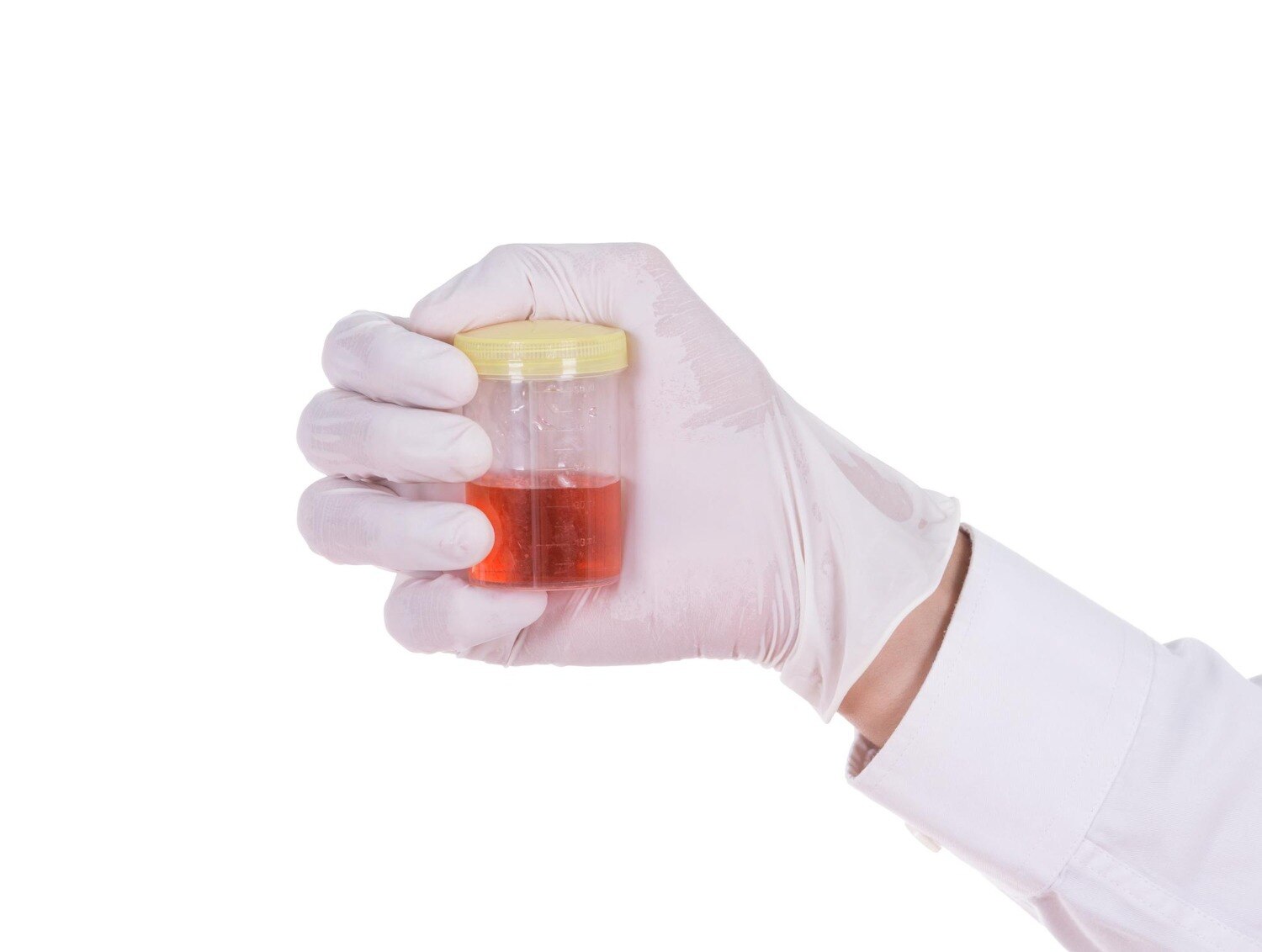
Red, pink, tea-coloured, or cola-coloured urine is visible blood in urine, also called gross hematuria. It needs medical evaluation even when the bleeding is painless, happens once, or disappears. No pain does not rule out a stone, infection, prostate condition, or urinary-tract cancer.
Microscopic hematuria is more than 3 red blood cells per high-power field on microscopy from one properly collected urine sample. A positive dipstick for blood is not a diagnosis; it can detect blood-related pigments and must be confirmed with microscopic examination.
Seek emergency care immediately if you have:
- Inability to pass urine
- Heavy bleeding or blood clots in urine; attention is required immediately because this is an urgent concern
- Severe flank or abdominal pain
- Fever or chills with burning, urgency, or other urinary symptoms
- Vomiting that prevents fluids or medicines
- Dizziness, fainting, or marked weakness
- A single functioning kidney, especially with pain, fever, or reduced urine output
Clots can block urine flow and cause painful retention or kidney damage. Fever with flank pain can signal an infected obstruction, which is a urological emergency. Even without these warning signs, visible blood requires prompt medical assessment rather than observation at home.
Could the blood have come from somewhere other than the urinary tract?
Red, pink, tea-coloured, or cola-coloured urine is not automatically urinary bleeding. Menstrual blood, vaginal bleeding, haemorrhoids, a cut on the genital skin, recent sexual activity, vigorous exercise, or trauma can place blood in or around the container and create blood in urine contamination.
1. Check the timing. A menstrual contamination urine sample or bleeding after sex should be repeated after the bleeding stops. A skin wound, haemorrhoid, exercise session, or minor trauma also warrants a new sample after the problem resolves.
2. Collect a clean-catch sample. Wash the genital area, begin urinating into the toilet, then collect the midstream urine in the sterile container. Tell the clinician about any vaginal bleeding, injury, exercise, or sexual activity when the sample was taken.
3. Do not diagnose infection from colour alone. Urinary tract infection hematuria should not be assumed without urinary symptoms and testing; when infection is suspected, obtain a urine culture and repeat urinalysis after UTI treatment, including microscopy.
Dehydration can concentrate urine and make it dark yellow, while beetroot can turn it pink or red. Neither explanation safely rules out true hematuria. If blood persists on a properly collected sample, seek risk-based evaluation rather than repeated antibiotic courses or continued attribution to the original explanation.
What happens during the first urological evaluation?
Your first hematuria evaluation combines a focused history, examination, and basic tests. The clinician asks about urinary symptoms, previous stones or infections, smoking, occupational chemical exposure, pelvic radiation, recent exercise or trauma, relevant family history, and the timing, amount, and colour of bleeding. Blood pressure measurement and a physical examination complete the initial assessment.
- Urinalysis with microscopy confirms red blood cells and can identify proteinuria, dysmorphic red cells, or cellular casts.
- Order a urine culture when infection is suspected, based on symptoms and examination; do not assume infection from blood alone.
- Check serum creatinine to assess kidney function. This serum creatinine hematuria assessment helps identify reduced filtration and changes the urgency of follow-up.
Review every medicine, including warfarin, apixaban, rivaroxaban, aspirin, and clopidogrel. Blood thinners and blood in urine are linked because these medicines can worsen bleeding or reveal an underlying urinary-tract lesion; they do not automatically explain hematuria. Never stop an anticoagulant or antiplatelet drug without instructions from the prescribing clinician.
Proteinuria, dysmorphic red cells, cellular casts, or reduced kidney function suggest a glomerular source and require nephrology referral. Nephrology assessment does not replace urological evaluation when kidney disease and a urinary-tract lesion could both be present.
When are ultrasound, CT, and cystoscopy needed?
Low or negligible-risk microscopic hematuria usually starts with repeat urinalysis within six months, not automatic cystoscopy and imaging. If blood persists, your risk category changes and further testing follows.
Intermediate-risk hematuria calls for cystoscopy plus renal bladder ultrasound. Ultrasound uses no radiation or iodinated contrast, but it is less sensitive than CT urography for some small renal masses, ureteral lesions, and urinary stones.
| Risk level | Recommended evaluation | What it shows |
|---|---|---|
| Low or negligible | Repeat urinalysis within six months | Confirms whether blood persists |
| Intermediate | Cystoscopy plus renal bladder ultrasound | Bladder and urethra; kidneys and ureters |
| High | Cystoscopy plus axial upper-tract imaging | Direct bladder inspection and detailed kidney and ureter imaging |
High-risk hematuria requires cystoscopy plus axial upper-tract imaging. Multiphasic CT urography is preferred when suitable for CT urography hematuria evaluation; it uses contrast phases to assess the kidneys, ureters, and urinary tract. If CT contrast or radiation is unsuitable, use MR urography.
When both CT and MR urography are unsuitable, retrograde pyelography combined with noncontrast imaging or ultrasound is an alternative; a non-contrast CT kidney stones study is useful when stones are suspected.
Cystoscopy for hematuria directly inspects the urethra and bladder, while imaging examines the kidneys and ureters. Imaging cannot replace cystoscopy when cystoscopy is indicated. Urine cytology is reserved for selected situations, such as equivocal cystoscopy or concern for carcinoma in situ, not used as a routine substitute.
How can you prepare for a urological evaluation?
Bring your records and a clear timeline; this makes the hematuria diagnostic pathway faster and prevents repeat testing.
1. Pack the urine report, urine-culture results, and imaging discs or reports. Add a complete medication list with doses, including warfarin, apixaban, rivaroxaban, aspirin, or clopidogrel. Note whether menstruation, vaginal bleeding, haemorrhoids, a skin wound, recent sex, exercise, or trauma could have contaminated the sample.
2. Record when visible bleeding started, whether it was red, pink, tea-coloured, or cola-coloured, how long it lasted, and whether you saw clots or pain. Use these details when deciding when to see a urologist for hematuria.
3. Do not delay emergency care while gathering records. Seek immediate assessment for inability to pass urine, heavy bleeding or clots, severe pain, fever or chills with urinary symptoms, vomiting, dizziness, fainting, or weakness. Do not stop an anticoagulant or antiplatelet drug independently; contact the prescribing clinician.
A urology clinic such as Dr. Vivek Jadhav Laser Stone and Urology Clinic in Thane West can coordinate your history, examination, urine and kidney-function tests, imaging decisions, and cystoscopy referral or procedure when indicated. If cystoscopy is planned, ask whether you need preparation instructions, medication adjustments, or a urine test beforehand.
Visible hematuria warrants evaluation; microscopic hematuria warrants properly confirmed, risk-based follow-up; emergency symptoms require immediate assessment.
Related services
Laser and endoscopic treatment of kidney stone Laser and endoscopic treatment of kidney stone A complete endoscopic prostate surgery typically refers to a minimally invasive procedure known as transurethral resection of the prostate (TURP). View service → |
LASER AND COMPLETE ENDOSCOPIC TREATMENT OF URETERIC STONES LASER AND COMPLETE ENDOSCOPIC TREATMENT OF URETERIC STONES Ureteric or Ureteral stones are the kidney stones that get stuck in one or both ureters. View service → |
Frequently asked questions
When does blood in urine require emergency care?
Seek emergency care if bleeding includes clots, you cannot urinate, or you have severe side or abdominal pain, fever, vomiting, faintness, or heavy ongoing bleeding.
Could the blood have come from somewhere other than the urinary tract?
Yes. Menstrual or vaginal bleeding, rectal bleeding, and blood from a skin wound can appear to be in the urine. A clean-catch sample and examination help identify the source.
What happens during the first urological evaluation?
The clinician reviews the bleeding, pain, urinary symptoms, medicines, smoking history, infections, and family history, then performs an examination and orders urine and blood tests when appropriate.
When are ultrasound, CT, and cystoscopy needed?
Ultrasound can assess the kidneys, bladder, and urine remaining after urination. CT can identify stones, bleeding, and other urinary-tract abnormalities. Cystoscopy lets the urologist inspect the urethra and bladder directly.
How can you prepare for a urological evaluation?
Record the urine colour, timing, clots, pain, fever, and urinary symptoms. Bring a medicine list, note menstrual timing, and ask whether you need a full bladder or fasting for imaging.
Related services
LASER & COMPLETE ENDOSCOPIC TREATMENT OF BLADDER STONES LASER & COMPLETE ENDOSCOPIC TREATMENT OF BLADDER STONES Bladder stones are hard masses of minerals that form stones in bladder. View service → |
Bladder Cancer surgery and treatment Bladder Cancer surgery and treatment Bladder cancer surgery typically involves the removal of cancerous tissue from the bladder. View service → |
Andrology and Male infertility treatment Andrology and Male infertility treatment Andrology focuses on male reproductive health, including male infertility. View service → |