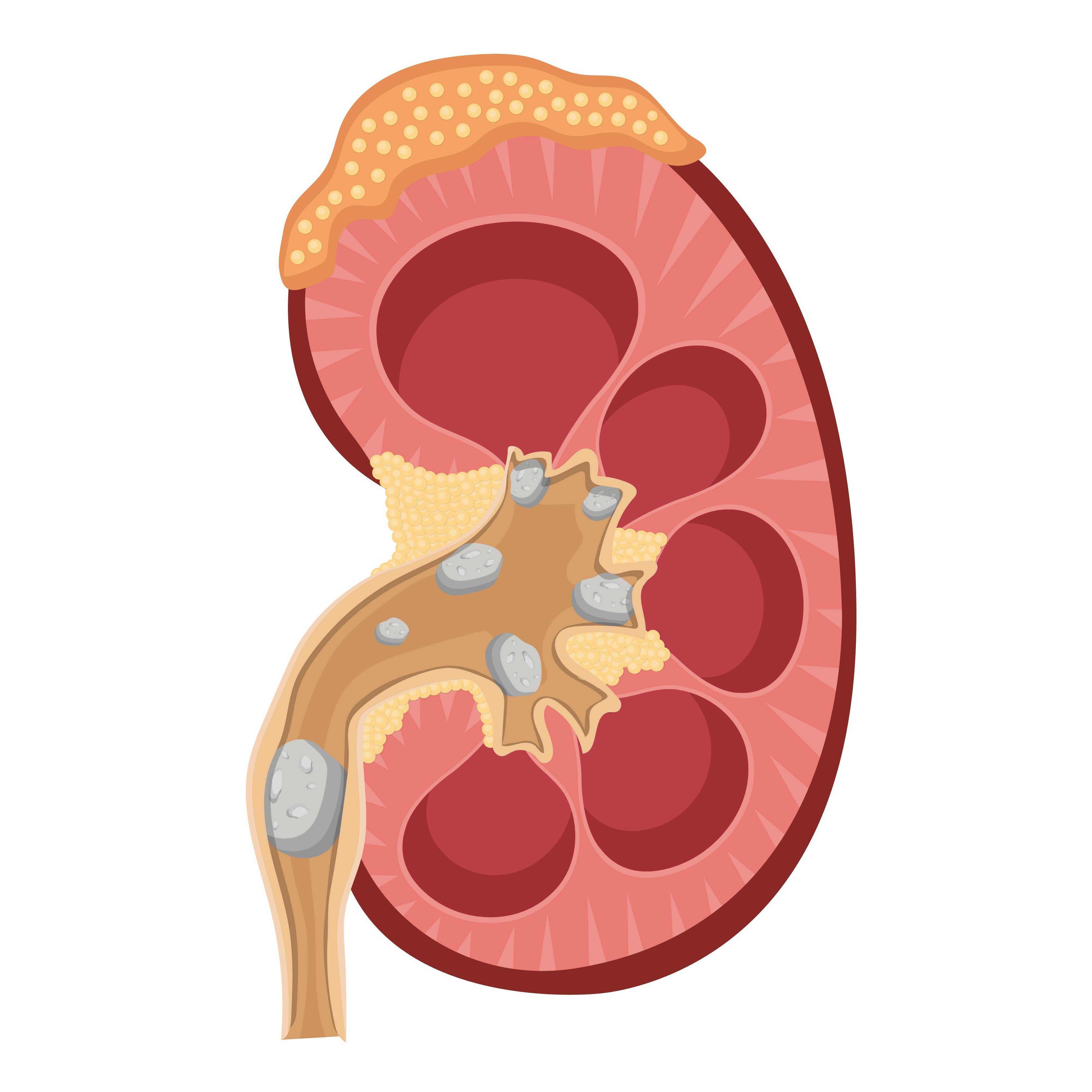
A 4 mm stone near the bladder is managed differently from a 4 mm stone near the kidney, and severe symptoms can change the plan even when the stone is small. By the end, you will know which scan findings, warning signs, size ranges, and treatment trade-offs clinicians use to decide between observation, medication, shock-wave treatment, laser ureteroscopy, and percutaneous nephrolithotomy.
Key takeaways
- Seek urgent care for fever, vomiting, inability to urinate, or uncontrolled pain.
- Use stone size, location, symptoms, and elapsed time to judge whether passage is likely.
- Choose removal by comparing stone burden, success, recovery, and procedure risks.
- Confirm stone clearance with follow-up imaging; pain relief alone does not prove removal.
First, determine whether the stone is safe to observe
Fever, chills, suspected infection, uncontrolled pain, repeated vomiting, or inability to urinate make a kidney stone emergency. A solitary kidney, pregnancy, or reduced kidney function also lowers the threshold for urgent assessment because obstruction can damage the only or already vulnerable functioning kidney.
An obstructed kidney infection is an emergency: treatment requires antibiotics and urgent drainage with either a ureteral stent or a percutaneous nephrostomy. Definitive stone removal waits until the infection resolves; a stent drains urine but does not remove the stone.
- Seek urgent medical care for fever or chills with stone pain, worsening or uncontrolled pain, repeated vomiting, inability to keep fluids down, inability to urinate, or new weakness.
- Ask for prompt review if you have one kidney, are pregnant, or have reduced kidney function. Pregnancy usually calls for ultrasound first because it uses no radiation; urgency still depends on infection, obstruction, symptoms, and kidney function.
- A non-contrast CT scan establishes kidney stone size and location, whether the stone is in the kidney or ureter, obstruction, hydronephrosis, stone density, and relevant anatomy. It is the clearest test when treatment planning needs precise measurements.
- A plain X-ray shows only radiopaque stones and is less precise for size and obstruction. Ultrasound can show hydronephrosis and some stones but may miss small ureteral stones, especially near the bladder.
Observation is reasonable only when symptoms are controlled, there is no infection or significant obstruction, and kidney function is stable. Follow-up imaging must confirm passage; pain stopping alone does not prove it.
Use size and time to judge passage before choosing removal
A practical kidney stone size treatment guide starts with diameter, not ends there. Stones under about 5 mm have the strongest case for observation when pain is controlled and there is no infection, significant obstruction, or worsening kidney function.
Stones around 5–10 mm have a less predictable stone passage likelihood; selected patients may try medication-assisted passage or choose active treatment. A ureteral stone above 10 mm is less likely to pass and is commonly discussed for removal.
| Size | Usual discussion | What changes the decision |
|---|---|---|
| Under 5 mm | Observation | Symptoms, obstruction, infection, kidney function, and follow-up imaging |
| 5–10 mm | Selected medical expulsive therapy or active treatment | Location, time since diagnosis, pain, and passage progress |
| Over 10 mm in the ureter | Removal is commonly discussed | Persistent obstruction, anatomy, density, and patient priorities |
An alpha-blocker kidney stone plan has its clearest role for a selected distal ureteral stone, particularly one over about 5 mm, near the bladder. Medical expulsive therapy distal ureter stone treatment is not appropriate when infection or urgent obstruction requires drainage.
Use an NSAID for renal-colic pain when medically safe, add prescribed anti-nausea treatment when vomiting interferes with fluids or medicines, and drink normally rather than forcing large volumes. Time matters: a stone still present after weeks, ongoing obstruction, or repeated pain deserves reassessment rather than indefinite waiting. Observation means planned imaging, not forgetting the stone.
Location can matter as much as the measurement
A kidney stone can remain quiet, while a same-sized ureteral stone causes colic and obstruction. A stone at the ureteropelvic junction (UPJ), where the kidney meets the ureter, can block drainage and produce hydronephrosis; a stone near the bladder has a shorter route left to travel.
| Location | Passage and treatment implications |
|---|---|
| Kidney stone | Often monitored if small, stable, and asymptomatic; treatment depends on growth, anatomy, obstruction, and symptoms. |
| Upper ureter stone | Less likely to pass than a same-sized distal stone; upper ureter stone treatment may involve shock waves or ureteroscopy. |
| Middle ureter stone | Passage is intermediate; ureteroscopy or observation depends on size, symptoms, obstruction, and time. |
| Lower ureter stone near bladder | More likely to pass, especially near the ureterovesical junction; selected stones may suit an alpha-blocker if urgent drainage is not needed. |
A ureterovesical junction stone obstruction can still damage kidney function or cause infection, so proximity to the bladder does not make it automatically safe.
Lower-pole kidney stones create a separate problem: a long or narrow infundibulum and an unfavorable infundibulopelvic angle can leave shock-wave fragments trapped in the dependent calyx, shifting treatment toward ureteroscopy or percutaneous removal.
An asymptomatic kidney stone can be monitored while stable. Growth, obstruction, infection, symptoms, a high-risk location, safety-critical work, or anticipated treatment difficulty favors active removal. A middle ureter stone that remains lodged deserves reassessment even if pain fades.
Match the procedure to the stone burden and the trade-offs
The practical choice depends on more than diameter: hardness, location, anatomy, obstruction, symptoms, and how many procedures you are willing to undergo all matter. A discussion of shock-wave lithotripsy versus ureteroscopy should include these trade-offs:
| Option | Main advantage | Main limitation |
|---|---|---|
| Shock-wave lithotripsy | Breaks the stone without entering the urinary tract | Works less reliably for hard or dense stones, large stones, lower-pole stones, and unfavorable anatomy; fragments can remain and require repeat treatment |
| Ureteroscopy with laser | Usually gives many ureteral stones a higher chance of being stone-free after one procedure | Requires anesthesia and urinary-tract access; laser fragmentation is common, and a temporary ureteral stent can cause urgency, frequency, flank discomfort, or blood in the urine |
| Percutaneous nephrolithotomy | Removes a large kidney-stone burden through a small access tract in the skin | More invasive, with hospital recovery and bleeding or infection risks |
The percutaneous nephrolithotomy stone size threshold is about 20 mm: above that, it is generally preferred because shock waves or ordinary ureteroscopy may require several sessions. Stones around 10–20 mm need an individual choice based on density, anatomy, location, symptoms, and patient priorities.
At Dr. Vivek Jadhav Laser Stone and Urology Clinic, specialist assessment can compare these options rather than selecting a procedure from size alone. Ask specifically about laser ureteroscopy recovery, the chance of repeat treatment, and whether you need a temporary stent.
Related services
LASER AND COMPLETE ENDOSCOPIC TREATMENT OF URETERIC STONES LASER AND COMPLETE ENDOSCOPIC TREATMENT OF URETERIC STONES Ureteric or Ureteral stones are the kidney stones that get stuck in one or both ureters. View service → |
Laser and endoscopic treatment of kidney stone Laser and endoscopic treatment of kidney stone A complete endoscopic prostate surgery typically refers to a minimally invasive procedure known as transurethral resection of the prostate (TURP). View service → |
Confirm clearance instead of equating pain relief with a cure
Pain stopping does not prove passage: a stone can remain lodged while obstruction continues. Confirm clearance by capturing the stone or using follow-up imaging for a kidney stone—ultrasound, a plain X-ray for a radiopaque kidney stone, or repeat low-dose non-contrast CT when precise confirmation is needed.
Clinicians plan the timing around stone size and location, obstruction, kidney function, and symptoms. A small observed stone needs reassessment rather than forgotten; worsening pain, fever, vomiting, reduced urine, or declining kidney function requires earlier review.
After treatment, imaging and tests check residual fragments, hydronephrosis, kidney function, and infection. A stent drains urine but does not remove the stone; remove or exchange it according to the procedure and complication risk. Ask specifically about kidney stone stent removal before leaving the clinic.
Take this decision checklist to your consultation:
- What are the exact size in millimetres, location, and density?
- Is there obstruction or hydronephrosis?
- Is infection present or excluded?
- How is kidney function?
- What is the expected passage time?
- What follow-up imaging will confirm clearance, and when?
- Will anesthesia be needed?
- What is the repeat-procedure risk?
- What recovery effort, stent symptoms, or work restrictions should you expect?
- Which option best fits your personal priorities?
Frequently asked questions
When is a kidney stone unsafe to observe?
Fever, chills, suspected infection, uncontrolled pain, repeated vomiting, or inability to urinate require urgent assessment. A solitary kidney, pregnancy, or reduced kidney function also lowers the threshold for prompt care.
How do size and time affect the decision to remove a kidney stone?
Smaller stones have a better chance of passing, while larger stones are less likely to pass without treatment. Persistent obstruction, ongoing symptoms, or failure to pass within the expected period shifts the decision toward removal.
Why does a stone’s location matter as much as its size?
A stone’s position in the kidney or ureter affects whether it can move into the bladder, pass naturally, or be reached efficiently with a procedure. Two stones with the same diameter can therefore need different plans.
How is successful kidney stone treatment confirmed?
Follow-up imaging confirms whether the stone and obstruction have cleared. Feeling better is not enough because pain can stop while a stone remains or urine flow stays blocked.